Figure 5.
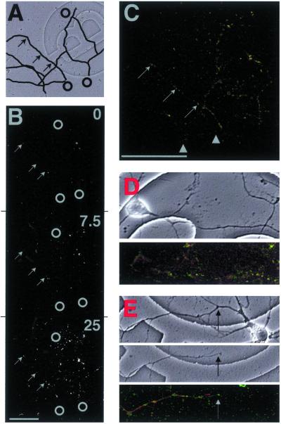
GFP hotspots colocalized with anti-ribosomal fluorescence. (A) Transmission image of isolated dendrites with black lines highlighting some larger dendrites. Black circles indicate sites of cell bodies before they were removed, and arrows show sites of example hotspots shown in B and C. One grid = 175 μm2. (B) MPLSM fluorescence images of dendrites in A after transfection with GFP mRNA and stimulation with DHPG for 7 min 30 s and 25 min. Gray circles indicate sites of cell bodies before they were removed, and arrows show example hotspots corresponding to those in C. (C) Confocal image of fluorescent dendrites from B that were fixed and counterstained with monoclonal anti-ribosomal Ab (red). Some red puncta appeared devoid of green fluorescence (arrowheads), but all green hotspots contained some red anti-ribosomal fluorescence (examples shown by arrows). [Bar = 100 μm.] (D) Example of an intact neuron stained under the same procedure as isolated dendrites in B and C. (Upper) Transmission image of neuron on gridded coverslip. One grid = 175 μm2. (Lower) Confocal image of green GFP fluorescence and red anti-ribosomal fluorescence. Intact dendrites showed the same fluorescence pattern as isolated dendrites in C. (E) Example of an isolated dendrite counterstained with tetramethylrhodamine B isothiocyanate (TRITC)-phalloidin (indicative of F-actin) under the same procedure as isolated dendrites in B and C (i.e., fluorescence pattern comparison using a protein not associated with translation). (Top) Transmission image of neuron on gridded coverslip before removal of cell body. (Middle) Transmission image of isolated dendrite after removal of the cell body. One grid = 175 μm2. (Bottom) Confocal image of green GFP fluorescence and red TRITC-phalloidin fluorescence. Arrow shows sever-point where cell body was removed. Many GFP hotspots did not contain red fluorescence; the pattern of fluorescence was different from the anti-ribosomal staining in C and D.

