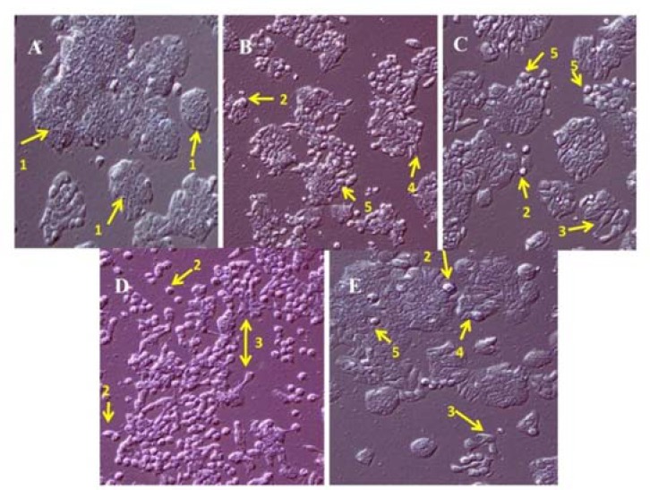
Fig. 1.

Morphology of HT-29 cell line by phase contrast microscopy at ×10. (A) untreated, (B) treated with concentration less than IC50 (75% of IC50) of methanolic extract of Stachys plifera, (C) alkaloid fraction, (D) terpenoid fraction, and (E) cisplatin after 24 h. The arrows indicate (1) normal cell, (2) apoptotic bodies, (3) cell debris, (4) cell shrinkage, and (5) detached cells.
