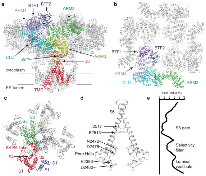
Figure 1. Structure of human type 3 IP3R in a ligand-free state.
a, Structure of hIP3R3 viewed in the plane of the membrane with the cytoplasmic domain at the top. A single subunit is colored according to domain with BTF1 in purple, BTF2 in blue, ARM1 in violet, the CLD in cyan, ARM2 in green, ARM3 in yellow, the JD in orange and the TMD in red. Gray lines represent the approximate position of the membrane. b, Structure of the cytoplasmic domain viewed from the cytoplasm. A single subunit is colored according to domain. ARM3 is removed for clarity. c, Structure of the transmembrane domain viewed from the cytoplasm. S1–S4 helices are colored red, S1′ and S1″ blue and S5, S6 and pore helix green. d–e, Structure of the ion conduction pathway viewed in the plane of the membrane and plot of pore radius. Residues comprising the luminal vestibule, the selectivity filter and S6 gate are highlighted. Front and rear subunits are removed for clarity.

