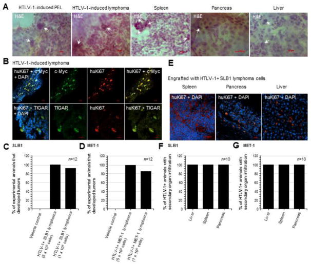
Fig. 6.
The TIGAR protein is highly expressed in tumor tissues with oncogene dysregulation in animals engrafted with HTLV-1-transformed SLB1 or MET-1 lymphoma T-cells. (A) Tissue sections were prepared from HTLV-1-induced tumors (PEL and solid lymphomas) and affected secondary tissues (spleen, pancreas, and liver) which were then stained with hematoxylin and eosin (H&E) to detect tumor cells with abnormal polylobate or pleomorphic nuclei and a large nuclear-to-cytoplasmic ratio (arrows in micrographs). (B) The HTLV-1+ lymphoma tumor tissues were immunostained with primary antibodies that recognize the human proliferating antigen Ki67 (huKi67; red signal), c-Myc oncoprotein (green signal), and human TIGAR protein (green signal) and the expression of these factors was analyzed by confocal microscopy. DAPI nuclear-staining (blue signal) was included to visualize the surrounding murine cells. (C and D) Graphs depicting the relative percentages of experimental animals, engrafted with 5×10⁵ or 1×10⁶ of the HTLV-1-transformed SLB1 or MET-1 cells, that developed tumors after 12-weeks compared to the Vehicle control group (n=12 animals per sample group). (E) Representative images showing the infiltration of secondary tissues (i.e., spleen, pancreas, and liver) by huKi67-positive (red signal) HTLV-1-transformed SLB1 tumor cells. DAPI nuclear-staining (blue signal) is provided for reference and the visualization of murine stromal cells. (F and G) Graphs depicting the percentages of HTLV-1+ engrafted animals which exhibited the infiltration of secondary tissues by tumor cells, based upon H&E-staining and/or Anti-huKi67 immunostaining results as shown in E (n=10 animals). All of the data in A, B, E, F, and G are representative of at least three sections for each tumor or secondary tissue analyzed.

