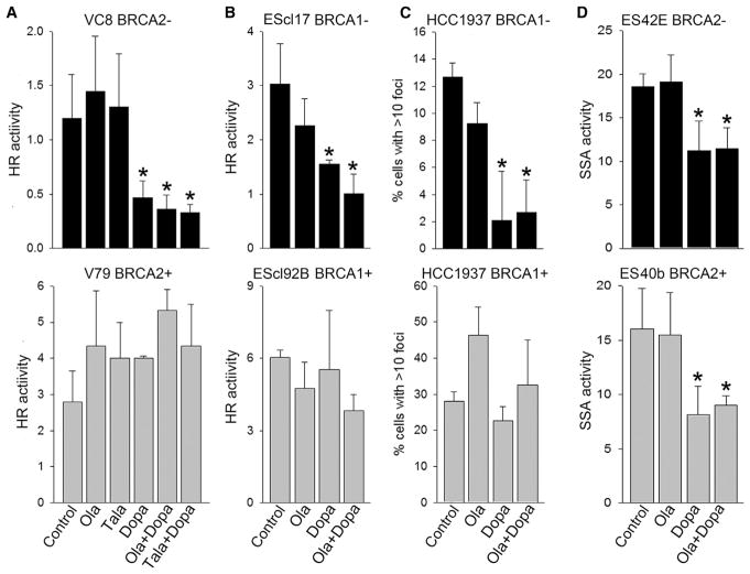
Figure 1. RAD52 Inhibitor 6-OH-Dopa Attenuated HR and SSA in BRCA1/2-Deficient Cells Treated with PARP Inhibitor Olaparib.
(A and B) BRCA2-mutated VC8 cells (BRCA2−) and BRCA2 wild-type V79 cells (BRCA2+) (A) and BRCA1−/− murine ES clone 17 cells (BRCA1−) and BRCA1 wild-type clone 92B cells (BRCA1+) (B) carrying DR-GFP cassette were co-transfected with I-SceI and DsRed cDNAs, followed by treatment with 5 μM olaparib (Ola), 50 nM talazoparib (Tala), and/or 10 μM 6-OH-dopa (Dopa), or were left untreated (Control). Results represent mean percentage of GFP+DsRed+ cells in DsRed+ population ± SD from three independent experiments; *p < 0.05 in comparison with untreated control.
(C) BRCA1-mutated HCC1937 cells (BRCA1−) and HCC1937 expressing wild-type BRCA1 (BRCA1+) were treated with 3 μg/mL cisplatin (Control) and cisplatin combined with 5 μM olaparib (Ola) and/or 10 μM 6-OH-dopa (Dopa). Results represent percentage of cells with more than ten RAD51 foci from three independent experiments (100 cells/experiment were evaluated); *p < 0.05 in comparison with untreated control.
(D) BRCA2−/− murine ES clone 42E cells (BRCA2−) and BRCA2 wild-type clone 40b cells (BRCA2+) carrying SA-GFP cassette were co-transfected with I-SceI and DsRed cDNAs, followed by treatment with 1.25 μM olaparib (Ola) and/or 20 μM 6-OH-dopa (Dopa), or were left untreated (Control). Results represent mean percentage of GFP+DsRed+ cells in DsRed+ population ± SD from three independent experiments; *p < 0.05 in comparison with untreated control.
See also Figure S1.

