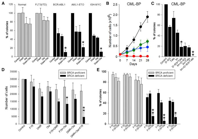
Figure 3. A Combination of PARP1 and RAD52 Inhibitors Exerted Stronger Synthetic Lethality Than Individual Inhibitors against Primary Cells from BRCA1/2-deficient Hematopoietic Malignancies.
(A) Lin−CD34+ cells from healthy donors (n = 4–8), BRCA-proficient FLT3(ITD)-positive AML (n = 3 or 4), and from BRCA1-deficient BCR-ABL1-positive CML-CP (n = 3–5), BRCA2-deficient AML1-ETO-positive AML (n = 3–6) and BRCA2-deficient IGH-MYC-positive BL cells were treated with 2.5 μM olaparib (Ola) and/or 2.5 μM Dopa.
(B) BRCA1-deficient BCR-ABL1-positive CML-BP cells were left untreated (black) or treated with 2.5 μM olaparib (blue), 20 μM Dopa (green), and olaparib + Dopa (red) for 28 days. Mean cumulative number ± SD of viable cells.
(C) CML-BP cells were treated with 2.5 nM talazoparib (Tala), 2.5 μM D-I03, and/or 1 μM imatinib (Im).
(D) BRCA pathway-deficient and BRCA pathway-proficient AML (n = 3 of each) were treated with 0.125 μg/mL daunorubicin (DNR), 1.25 μM olaparib, and/or 1.25 μM F79.
(E) BRCA pathway-deficient and BRCA pathway-proficient t-MDS/AML (n = 3 of each) were treated with 1.25 μM olaparib and/or 1.25 μM F79.
Cells were treated at 0 and 48 hr, followed by counting in trypan blue (B) or plating in methylcellulose at 96 hr (D); colonies were counted after 7–10 days (A, C, and E). Results represent mean percentage ± SD of surviving colonies/cells. *p < 0.05 in comparison with single and dual treatments, respectively, using Student’s t test; **p = 0.02 compared with DNR+Ola, DNR+F79, Ola, and F79 using the response additivity approach.

