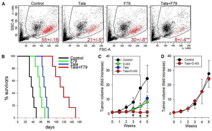
Figure 5. The Effect of a Combination of PARP1 and RAD52 Inhibitors against BRCA1-Deficient Primary AML Xenograft in NSG Mice and against BRCA1-Deficient Solid Tumor Growth in Nude Mice.
(A and B) NSG mice were inoculated i.v. with 106 BRCA1-deficient primary AML xenograft cells. One week later, the animals were treated with vehicle (Control), F79 aptamer (F79), talazoparib (Tala), or F79 + Tala (six mice per group) for 7 consecutive days.
(A) Representative plots of peripheral blood leukocyte (PBL) from treated mice; mean percentage ± SD of human CD45+ AML cells in peripheral blood leukocytes 3 weeks after leukemia injection; *p ≤ 0.005 and **p < 0.001 in comparison with Control and single-compound treatment, respectively, using Student’s t test.
(B) Kaplan-Meier survival curves.
(C and D) Nu/nu mice were inoculated s.c. with 106 BRCA1-deficient MDA-MB-436 cells (C) and BRCA1-proficient MDA-MB-436 +BRCA1 cells (D). Tumor-bearing animals were treated with D-I03, talazoparib (Tala), or D-I03+Tala for 7 consecutive days (three to five mice per group). Results represent mean ± SD fold increase of tumor volume; *p < 0.05 compared with individual agents.
See also Figures S5–S7 and Table S1.

