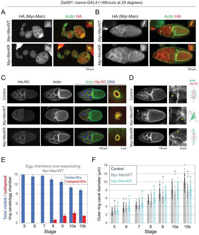
Figure 4. Expression of a membrane-tethered form of Misshapen leads to defects in ring canal stability and nurse cell fusion.
(A,B) Fluorescence images of egg chambers expressing Myr-HA-MsnWT or Myr-HA-MsnKR stained with an HA antibody. In addition to the ring canals and nurse cell membranes, Myr-HA-MsnWT and Myr-HA-MsnKR localize to punctae in the nurse cells and oocyte (arrowhead). Arrow in (B) points to a collapsed ring canal in a stage 10 egg chamber expressing Myr-HA-MsnWT. Boxes indicate regions of magnification. For magnified regions, scale bars are 2μm. (C,D) Fluorescence images of stage 10 control, Myr-HA-MsnWT, or Myr-HA-MsnKR expressing egg chambers. Boxes indicate regions of magnification in panels on the right. White arrowhead in (D) points to large actin bundle. Yellow arrowheads point to actin structures shown in the cartoons to the right. (E) Average number of visible and collapsed ring canals in egg chambers expressing Myr-HA-MsnWT. n=8–17 egg chambers/stage. Error bars are SEM. (F) Scatter plot showing the outer diameter of individual ring canals connecting nurse cells in control, Myr-HA-MsnWT, and Myr-HA-MsnKR expressing egg chambers. Bars indicate the average outer diameter at each stage. n = 44–159 ring canals/stage for each condition. Crosses were set up with the Gal80ts;nanos-GAL4 driver. Asterisks indicate significant difference compared to control (p<0.05, 2-tailed t-test).

