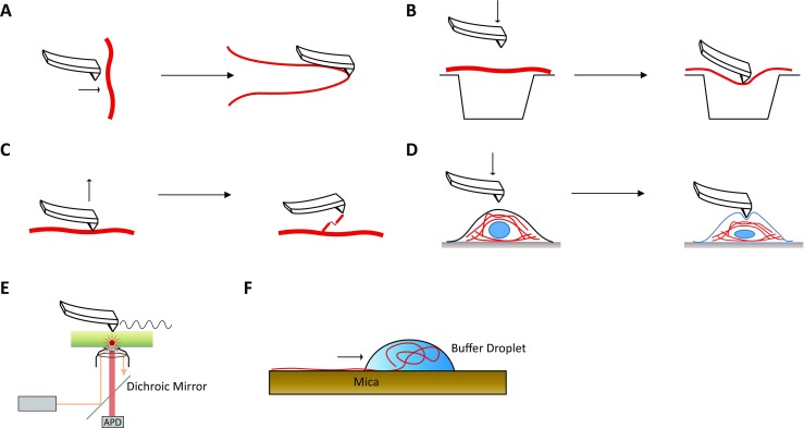
Fig. 5.

Overview of biophysical approaches to investigate the nanomechanical and structural properties of different desmin filaments. a Longitudinal stretching of isolated desmin filaments using the tip of an atomic force microscopy (Kreplak and Bar 2009). b Lateral stretching of IFs by pushing desmin filaments into small holes using the tip of an atomic force microscope (Guzman et al. 2006). c Molecule force spectroscopy using an atomic force microscope (Kiss et al. 2006). d Cellular stretching of transfected cells expressing mutant desmin (Plodinec et al. 2011). e Schematic overview of apertureless scanning near-field optical microscopy (aSNOM) (Harder et al. 2013). f Longitudinal stretching of desmin filaments using the withdrawing meniscus of a buffer droplet. Centrifugation is used to apply centrifugal forces (Kiss and Kellermayer 2014)
