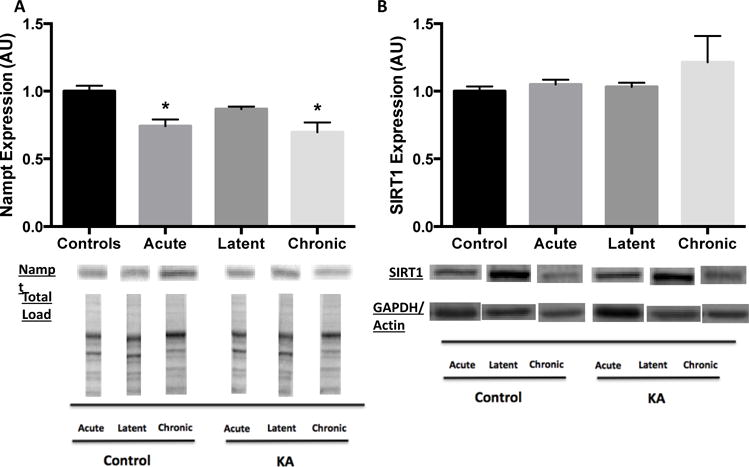
Figure 3.

Decreased Nampt protein expression during acute and chronic phases and no change in SIRT1 protein expression at all time points of epileptogenesis in hippocampus. Hippocampal nuclear-cytoplasmic (A) Nampt and (B) SIRT1 protein expression during epileptogenesis. Nampt expression normalized to total protein load for both Control and Kainic acid (KA)-treated at all time points. SIRT1 expression normalized to same loading control for both Control and KA-treated at each time point (acute: GAPDH, latent: GAPDH, chronic: actin). Data are normalized to Control mean value. Representative blot images below. Values are mean ± SEM (n = 3-14 per group). *, p < 0.05 vs Control.
