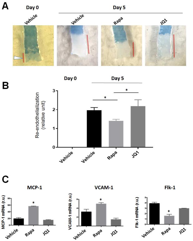
Figure 3. Effects of drug-loaded biomimetic nanoclusters on re-endothelialization in balloon-injured arteries.
Injections of biomimetic nanoclusters were performed as described in the Methods section. Animals were euthanized at 5 days post-angioplasty. Carotid arteries were collected and stained with Evans Blue.
A. Representative images of Evans Blue staining of carotid arteries. Denuded areas were stained blue; the unstained artery length is indicated by a red bar. Day-0 arteries were stained immediately after balloon angioplasty injury, thus there was no re-endothelialization at this time point.
B. Quantification of re-endothelialization. In every rat carotid artery, balloon angioplasty commonly left the proximal end uninjured (see arrowhead in A, Day-0). The length of this uninjured segment was therefore used as a common denominator to normalize unstained artery lengths in all conditions. Re-endothelialization was then calculated by subtracting the normalized value of the Day-0 condition in which no re-endothelialization occurred. Mean ± SEM, n=3 rats; *p<0.05.
C. Analysis of endothelial cell markers. Evans Blue-stained arteries were transiently fixed with RNAlater solution and subjected to RNA extraction and qPCR analysis. Quantification: Mean ± SEM, n=3 rats; *p<0.05 compared to vehicle control.

