
Extended Data Figure 2. FTCD depletion enables cancer cells to maintain THF pools and nucleotide synthesis even when treated with methotrexate (part 1).
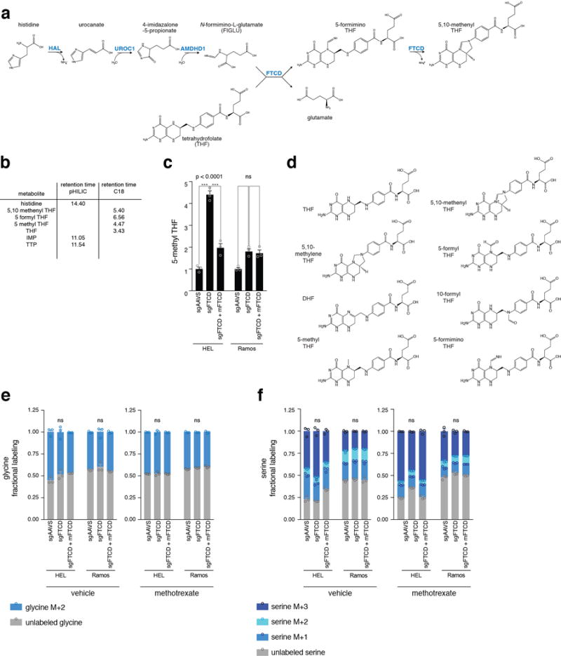
a. The histidine degradation pathway as previously described14,17–19. Enzymes are marked in blue. b. Metabolites detected by LC/MS and their corresponding retention times. The retention time for each detected metabolite is listed. The LC column used for the detection of each metabolite is also indicated. c. Greater pool of 5-methyl THF in vehicle-treated HEL cells following FTCD depletion. It is not readily clear why, in vehicle-treated HEL cells, FTCD depletion caused a reduction in THF levels (Fig. 2e). However, 5-methyl THF levels in these cells were increased significantly (Extended Data Fig. 2c and 3c), indicating that there was no depletion in the overall recyclable amounts of THF in these cells. 5-methyl THF levels were measured by LC/MS in vehicle-treated HEL and Ramos cells. 5-methyl THF levels were normalized to aminopterin as an internal standard. p-values were calculated using one-way ANOVA. n=3, biological replicates. d. Chemical structures of the folate entities found in cells and mentioned in Fig. 2c. e-f. Labeling rate by [U-13C] serine is not different between FTCD-depleted cells and control cells. Fractional labeling of glycine (e) and serine (f) is unchanged by FTCD depletion in HEL and Ramos cells in vehicle-and methotrexate-treated cells. Glycine and serine levels were normalized to isotopically-labeled glutamate as an internal standard. p-values were calculated for the unlabeled fraction by one-way ANOVA. n=3, biological replicates.
Source data for Fig. 2 and Extended Data Fig. 2 and 3 can be found in the file Source Data_2.
Abbreviation: UROC1 – urocanate hydratase 1.