
Extended Data Figure 6. In vivo histidine supplementation increases flux through the histidine degradation pathway and sensitizes tumors to methotrexate (part 1).
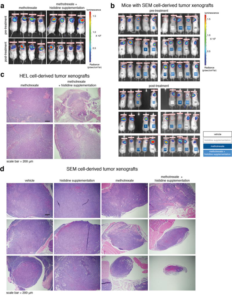
a. In vivo imaging of luciferase-expressing HEL tumor xenografts. Mice were imaged before (top images) and after (bottom image) five days of methotrexate treatment alone or in combination with histidine supplementation. For panels a,c, HEL cell-derived tumor-bearing mice: vehicle n=5, histidine supplementation n=4, methotrexate n=6, methotrexate + histidine supplementation n=6. Four mice per group are presented. b. In vivo imaging of luciferase-expressing SEM tumor xenografts. Mice were imaged before (top images) and after (bottom image) five days of treatment. All mice are presented. Mice are numbered in color by their experimental group. For panels b,d, SEM cell-derived tumor-bearing mice: vehicle n=6, histidine supplementation n=7, methotrexate n=7, methotrexate + histidine supplementation n=7. c. Additional images of H&E analyses of HEL cell-derived tumor sections from methotrexate-treated and methotrexate plus histidine supplementation-treated mice. d. H&E images of SEM cell-derived tumor sections from all treatment groups. Three mice per group are presented.
Source data for Fig. 4 and Extended Data Fig. 6–8 can be found in the file Source Data_4.