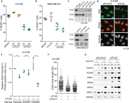
Fig. 1. TLK2 is required for efficient DNA replication.

(A) U-2-OS cells were pulsed with BrdU 48 hours after transfection with siRNAs against TLK1 or TLK2. The percentage of S-phase cells was subsequently quantified by analysis of DNA content using propidium iodide (PI) and staining for BrdU. Means and SD from technical replicates performed in at least biological duplicate are shown. (B) The percentage of S-phase cells was quantified in MDA-MB-231 cells pulsed with BrdU 72 hours after transfection and analyzed as in (A). (C) Western blot analysis of TLK1 and TLK2 from whole-cell lysates of U-2-OS (top) or MDA-MB-231 cells (bottom) 48 or 72 hours after siRNA transfection, respectively. (D) Immunofluorescence analysis of EdU incorporation in U-2-OS cells pulsed with EdU 30 hours after TLK2 siRNA transfection. Representative images are shown with PCNA as a marker for S-phase cells. (E) DNA replication in complemented U-2-OS cells. U-2-OS cells stably expressing siRNA-resistant WT or KD TLK2 were analyzed as in (D). See fig. S1F for Western blots for TLK2-WT or KD. EdU average intensities relative to parental cells from n = 3 independent biological replicates are shown with means and SD. One-sample and unpaired two-tailed t tests were used for statistical analysis of parental U-2-OS cells and complemented cell lines (TLK2WT and KD), respectively. **P < 0.01; n.s., not significant; a.u., arbitrary units. (F) Analysis of replication fork speed by DNA combing analysis. Length of CldU-labeled tracks (n > 250) was measured. One representative experiment of two biological replicates is shown, and median is indicated by a red line. (G) Analysis of replication factor chromatin loading in U-2-OS cells treated with or without UCN-01 30 hours after transfection. Cells were preextracted, and the chromatin pellet was subjected to Western blotting. One representative experiment of two biological replicates is shown.
