. 2018 Jul 31;7(2):119–128. doi: 10.7774/cevr.2018.7.2.119

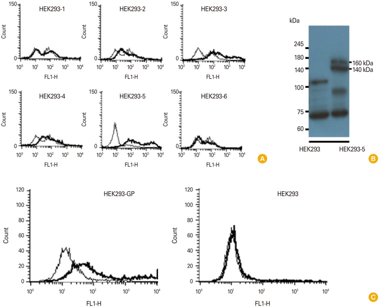
Fig. 1. Stable transfection of HEK293 cells with pcDNA3-GP and selection of GP-expressing HEK293 cells (A, B), and evaluation of sera obtained after GP DNA vaccination for their ability to recognize GP expressed on the cell surface. (A) HEK293 cells were stably transfected with pcDNA3-GP. The G418-selected cell colonies (HEK293-1, 2, 3, 4, 5, 6) were reacted with 2 µL of GP-specific immune sera, followed by reaction with FITC-conjugated anti-mouse IgG (whole molecules) for flow cytometry. The cells were measured for reactivity to GP expressed on HEK293 cells. (B) HEK293-5 cells which prominently reacted with anti-GP sera were further cultured and then the cell lysates were run on a 10% SDS-PAGE gel, transferred to a nitrocellulose membrane and reacted with commercial anti-GP antibodies for Western blot assay. (C) Mice were injected by IM-EP with pcDNA3-GP (50 µg/mouse) at 0 and 4 weeks. The mice were bled at 2 weeks following the final injection and sera were collected. (C) The sera were reacted with 5×10⁵ HEK293-GP and control HEK293 cells, followed by incubating with FITC-conjugated anti-mouse IgG (Fc) for flow cytometry. GP, glycoprotein; FITC, fluorescein isothiocyanate; SDS-PAGE, sodium dodecyl sulfate polyacrylamide gel electrophoresis; IM, intramuscular; EP, electroporation.

Fig. 1