
Figure 2. CCR2- and CCR2+ cardiac macrophage populations are maintained through distinct mechanisms.
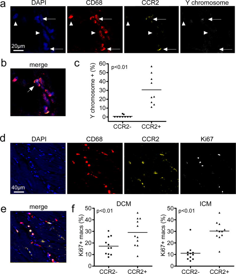
a, In situ hybridization and immunostaining of endomyocardial biopsy specimens obtained from recipients of sex mismatch heart transplants (n=9). All specimens were obtained from male patients who had received a heart from a female donor >1 year prior to biopsy. DAPI (blue), CD68 (red), CCR2 (yellow), and Y chromosome (white). Arrows: CCR2+ macrophages, arrowheads: CCR2- macrophages. b, Merged image from a. 400X magnification. Arrow denotes CCR2+ macrophage containing a Y chromosome. c, Percentages of CCR2- and CCR2+ macrophages that contain a Y chromosome (n=9). Each data point represents a biologically independent biopsy specimen and the line refers to the mean value. Mann Whitney test (two-sided), p<0.0001. d, Cell proliferation of CCR2- and CCR2+ macrophages, as assessed by immunostaining for CD68 (red), CCR2 (yellow), and Ki67 (white). Each data point represents a biologically independent heart failure specimen and the line refers to the mean value. Mann Whitney test (two-sided): DCM, p=0.0036 and ICM, p=0.006. e, Merged image from d. 200X magnification. f, Percentage of CCR2- and CCR2+ macrophages (macs) staining for Ki67 in hearts from DCM (n=11) and ICM (n=11) patients.