
Fig. 5. p190 controls Eg5activity.
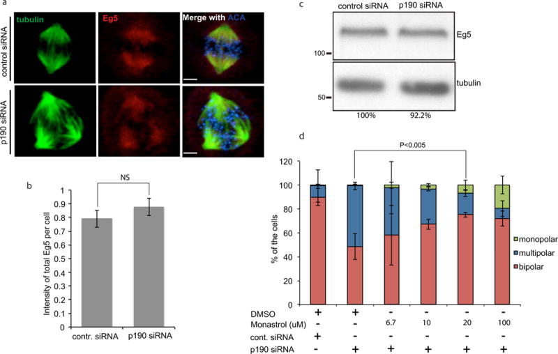
a. Eg5 localized properly in p190-depleted cells. Confocal images of control- and p190-siRNA treated cells that were fixed and stained with α-tubulin, ACA and anti-Eg5 antibody. Images shown are representative of n>20 bars represent 4 μm. b. Quantification of Eg5 intensity at poles in HeLa cells (p =NS). c. Immunoblot of endogenous Eg5 in control and p190 KD cells. Eg5:tubulin ratio were quantified and amount were reported by percentage. d. Multipolar spindles induced by p190 depletion are rescued by inhibition of Eg5 activity. p190-depleted and double thymidine-blocked cells were released from thymidine for 10 hours and treated with DMSO or 6.7μM, 10μM, 20μM or 100μM monastrol for one and a half hrs. Cells were fixed and stained with tubulin and ACA antibodies, and monopolar, bipolar and multipolar cells were quantified. Results are mean ± s.d from three independent experiments. The rescue of bipolar spindles by 20 uM monastrol is statistically significant (*P<0.005).