
Figure 2. Detailed analysis stage.
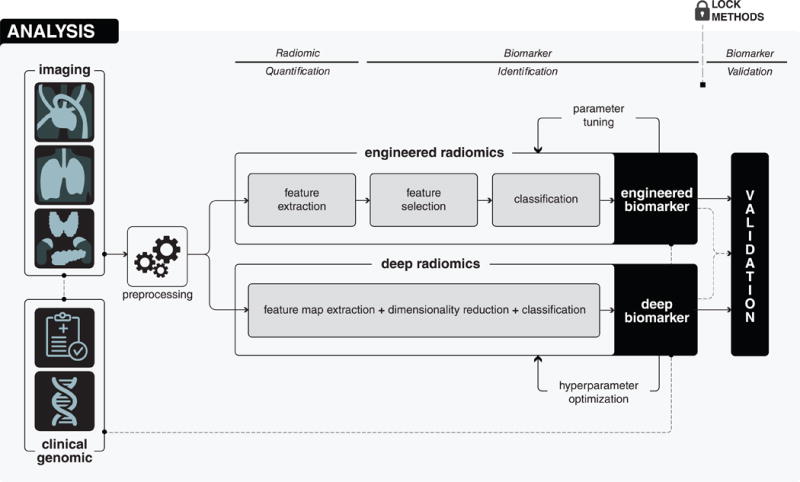
Analysis begins with the preprocessing of medical images in order to avoid different technical variability and batch effects. After that, in the radiomic quantification step, radiomic descriptors, capturing different phenotypic characteristics of diseased tissues are quantified. Radiomic quantification can either be done using engineered features or deep learning methods. According to the quantification method, in the biomarker identification step, appropriate analysis methods are explored and suitable methods are applied to develop biomarkers. Finally, in the biomarker validation step, the developed biomarker is validated in the locked and independent validation cohort.