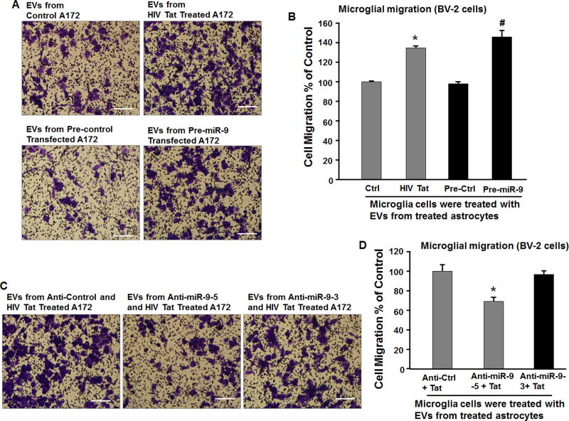
Figure 3. Increased migration of microglia treated with EVs released from Tat treated astrocyte cultures.
(A, C) The representative phase images of migratory BV-2 stained by crystal violet. Scale bar = 5 µm. (B) EVs isolated from Tat or pre-miR-9 treated A172 cells treatments significantly increased the number of BV-2 cells that were capable of migrating through the membrane of the inserts. * P < 0.05 versus control; # P < 0.05 versus Pre-Ctrl. (D) EVs isolated from Tat and anti-miR-9 treated A172 cells treatments significantly decreased Tat-induced migratory BV-2 cells. * P < 0.05 versus Anti-Ctrl.

