Figure 4.
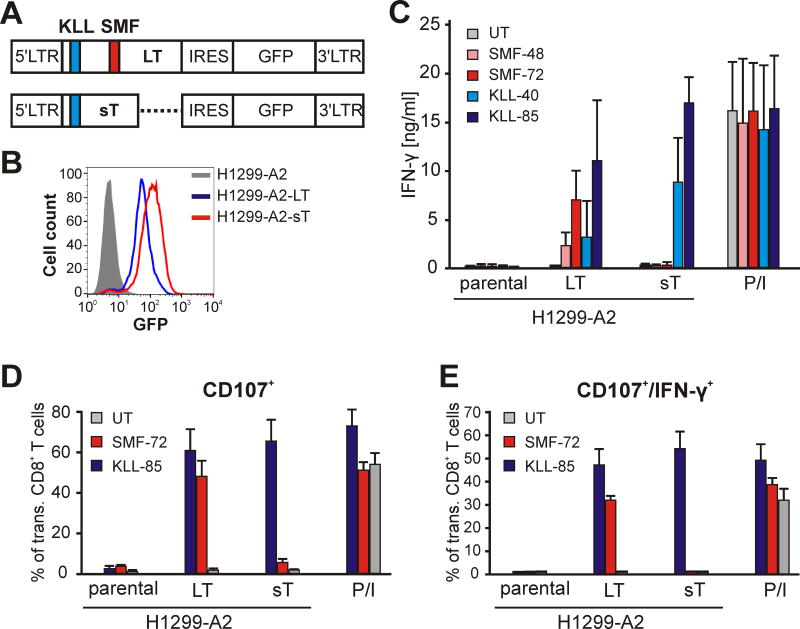
SMF- and KLL-epitopes are naturally processed. A. Schematic of MP71 vectors encoding LT or sT antigens as bicistronic constructs with GFP-expressing sequence segregated by internal ribosomal entry site (IRES). LTR: long terminal repeats. Positions of KLL (blue) and SMF (red) epitopes are indicated. B. Expression of LT (blue) and sT (red) antigens in H1299-A2-LT or H1299-A2-sT, respectively, cells as determined by fluorescence intensity of GFP. C. TCR-engineered human T cells as indicated were assayed for response in IFN-γ production after co-culturing with MCV-negative H1299-A2 cell line or derivatives expressing LT or sT antigens. TCR-independent stimulation of T cells with PMA/Ionomycin (P/I) served as positive control. Diagrams show means and standard deviations of 3 different experiments. D and E. Proportion of CD107+ or CD107/IFN-γ-double positive cells among SMF- or KLL-TCR expressing CD8+ T cells as indicator of cytotoxic activity after co-incubation with target cells as indicated. Diagrams show means and standard deviations of two experiments with T cells from different donors.

