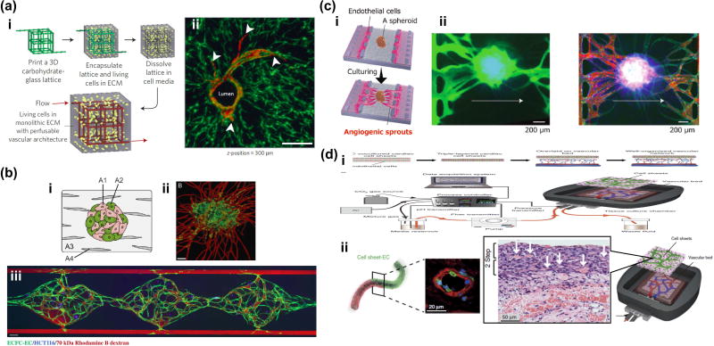
Figure 2.
Cutting-edge technologies to vascularize tissues in vitro. (a) Engineering of 3D vascular networks in extracellular matrix. (i) 3D casting mold was fabricated with carbohydrate glass lattice by 3D printer. After dissolving the lattice, endothelial cells were seeded into the 3D microchannels. (ii) Endothelialized microchannels and angiogenic sprouting with stromal cells. Potentially, this method could be applied to vascularization of various types of tissues, even relatively large tissues. (b) Vasculogenesis based vascularization in a microfluidic device. Flow of fluorescently tagged dextrans show the perfusability and connectivity of vascular networks to supply sufficient nutrients to HCT116 cells in a gel. This strategy is promising for high-throughput drug screening of vascularized micro-tissues (c) (i) Angiogenesis based vascularization of spheroid in a microfluidic device. (ii) Perfusion of fluorescently tagged dextrans showed that of angiogenic sprouts formed anastomoses with prevascularized networks inside the spheroid, preventing necrotic cell death in the core region. This method could be applied to vascularization of various types of spheroids and organoids for drug testing. (d) Vascularization of sheet-like tissues using an ex vivo vascular bed. Rat vascular bed was extracted and connected to a perfusion system in vitro. Stacked cell sheet with epithelial cells and endothelial cells were placed on this vascular bed. This ex vivo system has promise for the extension of culture time which is currently limited by the lack of vasculature (ii).

