
Fig. 3.
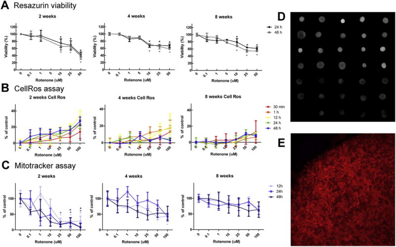
Rotenone produces different effects at different windows of exposure and preferentially affects dopaminergic neurons in BrainSpheres. Cells were exposed to different concentrations of rotenone for up to 48 h at 2, 4 and 8 weeks of differentiation. (A) shows cell viability at different concentrations of rotenone after 24 and 48 h exposure. (B) Reactive oxygen species (ROS) production after 0.5, 1, 12, 24 and 48 h exposure to different concentrations of rotenone. (C) Mitochondrial dysfunction after 12, 24 and 48 h rotenone exposure. D) Selected MitoTracker images. E) confocal image of a MitroTracker stained BrainSpheres.