
Fig. 4.
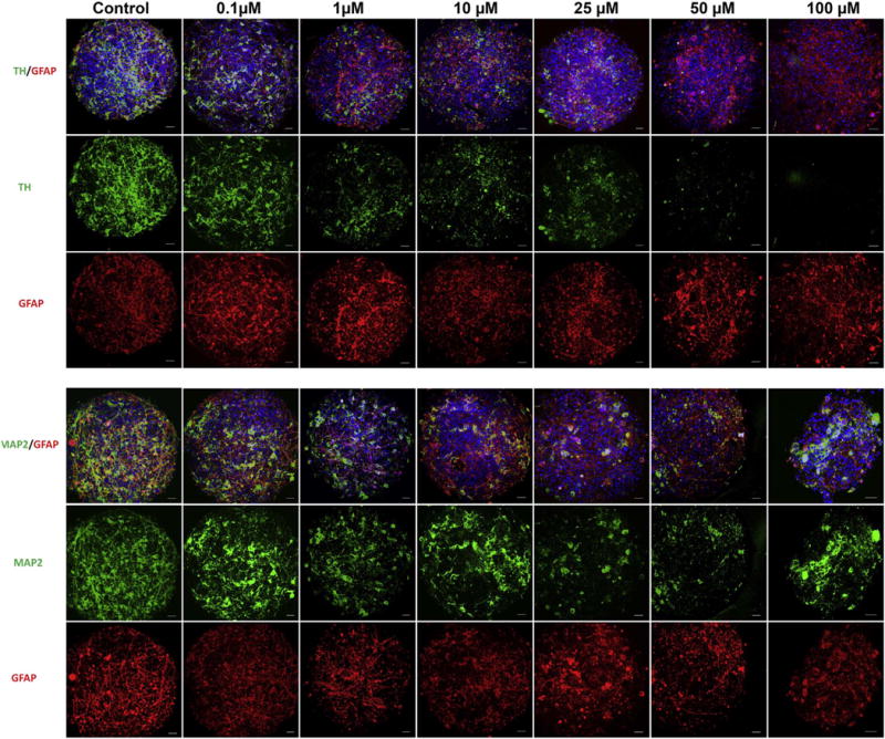
Immunohistochemistry of rotenone treated Human iPSC-derived BrainSpheres. BrainSpheres were collected after 8 weeks of differentiation and exposed to rotenone for 24 h. Confocal images were taken for neuronal (MAP2), astrocytic (GFAP) and dopaminergic neuronal (TH) cell populations in BrainSpheres exposed to increasing concentrations of rotenone. There was a progressive decrease in the overall neuronal population that appeared to be dopaminergic specific, as TH+ neurons were differentially affected with a relative preservation of MAP2+ neurons and GFAP+ astrocytes. Scale bar: 20 μm.