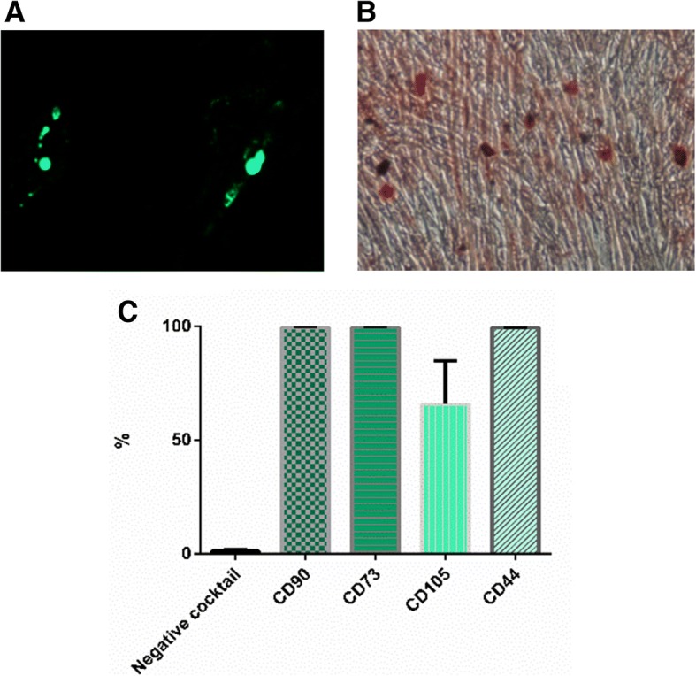
Fig. 1.

Characterization of UCMSCs. Mesodermic differentiation of UCMSCs. Clinical-grade UCMSCs differentiated into adipocytes (a) and osteocytes (b). Representative images are shown at × 10 magnification. Immunophenotypic analysis of UCMSCs by flow cytometry (c). UCMSCs presented the typical immunophenotype of MSCs. Negative cocktail includes CD34, CD45, CD11b, CD19, and HLA-DR markers. Results are shown as percentages of positive cells and are expressed as mean ± SEM. (n = 3)
