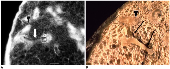
Fig. 4. Pathologic basis of tree-in-bud lesion.
Post-mortem radiograph (A) and gross photograph (B) of same specimen in patient with pulmonary tuberculosis. Impacted cheesy material within smooth-marginated bronchiole (arrows) continues to larger, rather ill-defined alveolar ducts (arrowheads). Bar indicates 1 mm. Reprinted with permission from authors' reference 1. Adapted from Im et al. Radiology 1993;186:653-660

