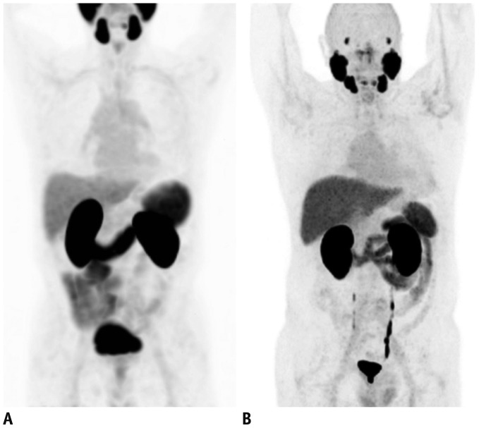
Fig. 1. Representative images of PSMA PET.
A. 68Ga-PSMA-11 PET shows normal biodistribution of PSMA across body; lacrimal and salivary glands, liver, spleen, kidneys, and intestines. B. 18F-DCFPyL PET demonstrates normal biodistribution of PSMA which is similar to 68Ga-PSMA-11 PET with better image resolution. These images were reprinted with permissions from reference articles 28 and 81, respectively. Adapted from Fendler et al. Eur J Nucle Med Mol Imaging 2017;44:1014-1024, 2017;44:2117-2136 [28] and Sheikhbahaei et al. Eur J Nucle Med Mol Imaging 2017;44:2117-2136 [81], with permissions of Springer Science and Bus Media B V. PSMA = prostate-specific membrane antigen

