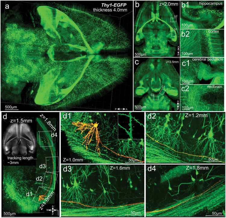
Fig. 6.
Whole-brain imaging and tracing of individual neurons and axons. Adult (60 days of age) Thy1-EGFP mouse brain was cleared following the PEGASOS passive immersion procedure. The dorsal-to-ventral thickness of the brain is around 4 mm after processing. a Whole-brain image acquired with a 10×/0.30 objective. Optical sections obtained at 2.0 mm and 3.5 mm are displayed in b and c. Boxed areas are enlarged to show hippocampus (b1), cortical neuron (b2), cerebral peduncle (c1) and midbrain (c2). Tracking individual axons in 3-D requires objective with higher NA to achieve high axial resolution. d Tiling images acquired with a 20×/0.95 objective on a confocal microscope show the tracking course of one neuron and its axon in 3-D. Inset at the corner was acquired with a 5× objective at the plane where the target neuron is located. Two arrows indicate the approximate beginning and ending positions of the tracking. Boxed areas are enlarged in panels d1 to d4. Boxed area in d1 is enlarged in the insert to show the dendritic spines. A, anterior; P, posterior; L, lateral; M, medial

