Figure 1.
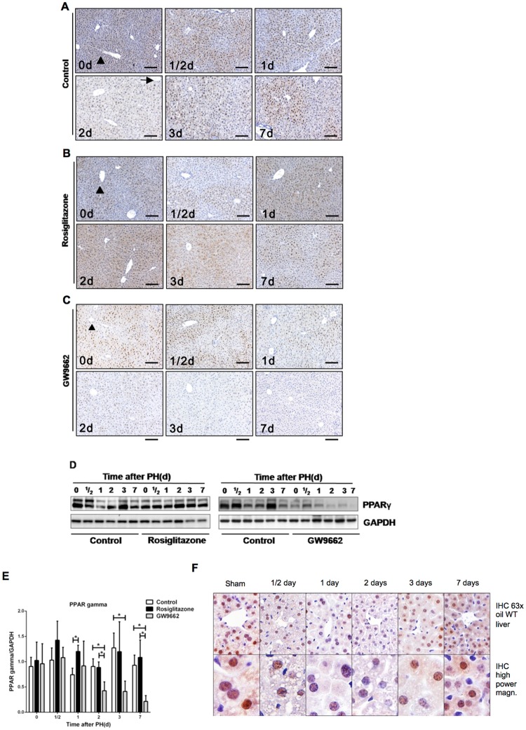
PPARγ inhibits hepatocellular proliferation during mouse liver regeneration (A) Hepatic PPARγ expression during mouse liver regeneration. Representative Immunohistochemistry (IHC) analysis for PPARγ in untreated mice, (B) rosiglitazone-treated mice, and (C) GW9662-treated mice at different time points after PH. Scale bar: 200 μm. Arrow: Centrilobular zone, Arrowhead: Periportal zone. (D) Hepatic expression of PPARγ protein in control mice, rosiglitazone-treated mice and GW9662-treated mice at different time points after partial hepatectomy. (E) Quantification of Western blots after analysis of three biological triplicates. (F) Analysis of cellular localization of PPAR gamma at different time points after PH in wild type mice. *p < 0.05.

