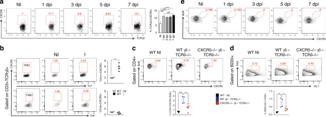
Fig. 4.
A subset of γδ T cells expresses CXCR5. a Frequency of γδ T cells expressing CXCR5 in the dLN of non-immunized (NI) WT mice and 1, 3, 5, and 7 days post CFA immunization (dpi) (n = 6 mice/group). b Frequency of Vγ1 and Vγ4 γδ T cells expressing CXCR5 from dLN of non-immunized (NI) WT mice and 3 days after CFA immunization (I) (n = 4 mice/group). c, d In vivo Tfh cell induction (c) and germinal center formation (d) in dLNs of TCRδ−/− mice transferred intravenously with 5 × 10⁵ γδ T cells from either WT or CXCR5−/− mice. Three days after transfer, recipient mice were immunized or not (WT NI) with CFA and 7 days thereafter sacrificed for flow cytometric analysis (n = 4–5 mice/group, where γδ T cells are sorted from a pool of 10 mice/group). e Representative FACS plots showing frequency of γδ T cells expressing CXCR5 and Bcl6 in dLN of non-immunized (NI) WT and 1, 3, 5, and 7 days post CFA immunization (dpi) (n = 5 mice/group/time point). These data are representative of at least two independent experiments (a, b, e). Data are shown as mean + SEM. One-way ANOVA was used. NS non-significant, *p < 0.05, **p < 0.01, ***p < 0.001, ****p < 0.0001

