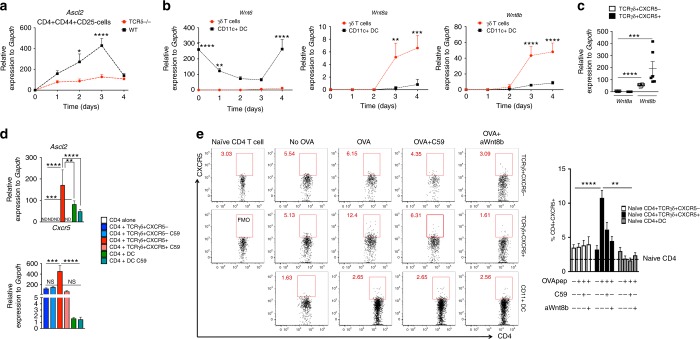
Fig. 7.
TCRγδ+CXCR5+ cells release Wnt ligands to induce CD4+CXCR5+ cells. a Quantitative RT-PCR analysis of Ascl2 mRNA from dLN-sorted CD4+CD44+CD25− T cells of WT and TCRδ−/− mice before immunization (day 0) and 1, 2, 3, and 4 days after CFA immunization (n = pooled cells from 5 mice/group/time point). Data are from three experiments. b Wnt6, Wnt8a, and Wnt8b from dLN-sorted γδ T cells and CD11c+ dendritic cells (DC) of WT mice before immunization (day 0) and 1, 2, 3, and 4 days after CFA immunization (n = pooled cells from 5 mice/group/time point). Data are from three experiments. c Wnt8a and Wnt8b from dLN-sorted TCRγδ+CXCR5− and TCRγδ+CXCR5+ cells of WT mice 4 days after CFA immunization (n = pooled cells from 5 mice/group). Data were combined from b. d Quantitative RT-PCR analysis of Ascl2 (36 h in co-culture) and Cxcr5 (72 h in co-culture) mRNAs from OT-II-Foxp3-GFP mouse-sorted naive CD4+ T cells cultured in the presence of OVA323–339-loaded TCRγδ+CXCR5−, TCRγδ+CXCR5+cells or CD11c+ DCs treated or not with the porcupine inhibitor Wnt-C59 (C59; 1 μM) for 3 days at 37 °C. γδ T cells and DCs were sorted from WT mice (pooled from 15 mice/experiment) 4 days after CFA immunization. Data are from three experiments. e CXCR5 induction on naïve CD4 T cells from OT-II-Foxp3-GFP mice co-cultured (3 days at 37 °C) with or without OVA323–339-loaded TCRγδ+CXCR5−, TCRγδ+CXCR5+ or CD11c+ DCs in the presence or not of either 1 μM of Wnt-C59 (C59) or 20 μg ml−1 of anti-Wnt8b monoclonal antibody (aWnt8b) from WT mice 4 days after CFA immunization (n = pooled cells from 15 mice/experiment). Data are from at least two experiments. Data are shown as mean + SEM. Two-way ANOVA (a, b) or one-way ANOVA (c–e) were used. ND not detected, *p < 0.05, **p < 0.01, ***p < 0.001, ****p < 0.0001

