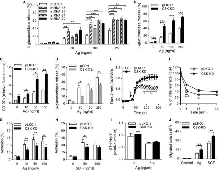
Figure 1.
Enhanced degranulation, calcium response, and chemotaxis toward antigen and SCF, but reduced adhesion to fibronectin and FcεRI–IgE complexes internalization in bone marrow-derived mast cells (BMMCs) with C-terminal Src kinase (CSK)-KD. (A) β-glucuronidase release was analyzed in BMMCs transduced with individual CSK-specific shRNAs (35, 36, 64, or 90) in pLKO.1 vector or empty pLKO.1 vector (pLKO.1). The cells were sensitized with IgE and activated with various concentrations of antigen (Ag) for 30 min. The data represent means ± SEM from 4–8 independent experiments performed in duplicates or triplicates. (B) β-glucuronidase release from BMMCs transduced with a pool of shRNAs (CSK-KD) or empty pLKO.1 vector was examined as in (A). The data represent means ± SEM from eight independent experiments performed in duplicates or triplicates. (C) Presence of CD107a on the cell surface in BMMCs with CSK-KD or empty pLKO.1 vector used as a control. The cells were sensitized with IgE, activated with various concentrations of antigen for 10 min and the presence of CD107a on the cell surface was analyzed by flow cytometry. The results shown are means ± SEM from three independent experiments performed in duplicates. (D) β-glucuronidase release from stable cell line derived from BMMCs transduced with empty vector (pCDH) or pCDH vector containing hCSK-mCherry construct (CSK-OE) was analyzed as in (A). The data represent means ± SEM from three independent experiments performed in duplicates or triplicates. (E) Calcium response examined in BMMCs with CSK-KD and corresponding controls. The cells were sensitized with IgE, loaded with Fura-2, and activated with antigen (100 ng/ml) added as indicated by an arrow. Data represent means ± SEM calculated from six independent experiments each performed in duplicate. (F) IgE internalization in BMMCs with CSK-KD or control pLKO.1 cells. The IgE-sensitized cells were activated with antigen (500 ng/ml) for various time intervals and fixed with 4% paraformaldehyde. IgE was quantified using AF488-labeled anti-mouse IgG (IgE cross-reacting) antibody by flow cytometry. Means ± SEM calculated from three independent experiments are shown. (G,H) Cell adhesion to fibronectin-coated surfaces. BMMCs with CSK-KD or pLKO.1 control cells were sensitized with IgE, loaded with calcein, and activated with various concentrations of antigen (G) or SCF (H). Fluorescence was determined before and after washing out non-adherent cells, and the percentages of adherent cells were calculated. The results indicate means ± SEM from five independent experiments. (I) The presence of β1-integrin on the cell surface in BMMCs with CSK-KD and corresponding control cells analyzed by flow cytometry. (J) BMMCs with CSK-KD or pLKO.1 control cells were sensitized with IgE and their migration toward antigen (250 ng/ml) or SCF (50 ng/ml) was determined. The data represent means ± SEM from five independent experiments each performed in duplicates. Statistical significance of intergroup differences was determined using one-way ANOVA with Tukey’s post-test (A) or unpaired two-tailed Student’s t-test (B–D,G–J) or two-way ANOVA with Bonferroni post-test (E,F). *P < 0.05; **P < 0.01; and ***P < 0.001.

