. 2018 Aug 7;59(7):816–826. doi: 10.3349/ymj.2018.59.7.816

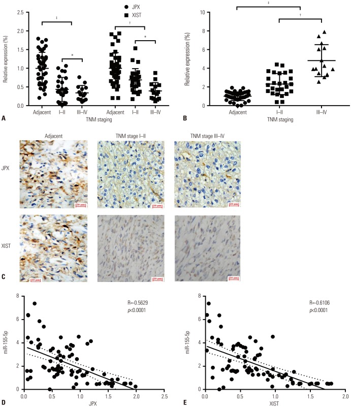
Fig. 1. XIST and JPX expression levels inversely correlated with HCC development and miR-155-5p expression level. (A and B) Expression levels of XIST and JPX significantly decreased in 20 HCC tissue specimens compared to adjacent counterparts and further decreased in advanced staged HCC tissue specimens, compared to early staged ones, while increases in miR-155-5p expression are significantly correlated with HCC oncogenesis and progression. Data were normalized to the mean of adjacent tissue and are presented as fold changes. (C) Representative images of JPX and XIST hybridization histochemistry in situ (magnification, ×200). Long non-coding RNAs in the frozen section were hybridized with biotin-labeled riboprobes in a complementary base pairing manner (pale brown). Cell nuclei were counterstained with hematoxylin (cyan). (D and E) Correlation analysis showing that expression levels of JPX and XIST are inversely correlated with those of miR-155-5p in all 20 pairs of HCC and adjacent tissue. Relative expression in (A) and (B) was used for correlation analysis in (D) and (E). *p<0.05, p<0.01, p<0.001. XIST, X-inactive specific transcript; JPX, just proximal to XIST; HCC, hepatocellular carcinoma.

Fig. 1