Figure 1.
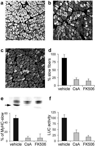
CsA and FK506 block the expression of MyHC-slow in regenerating soleus muscle. (a–d) Immunofluorescence analysis of sections stained with an antibody specific for MyHC-slow. Note that regenerating soleus muscles from rats treated with CsA (b) or FK506 (c) show a decreased proportion of reactive fibers compared with rats treated with vehicle (a). The percentage of fibers expressing MyHC-slow in the three experimental groups is shown in d. (e) SDS/PAGE profile of MyHCs (Upper). Note that the high mobility MyHC-slow band (arrow) is abundant in control muscle (left lane) but is barely detectable after treatment with CsA (center lane) or FK506 (right lane). This decrease is confirmed by quantitation of densitometric values of the percentage of MyHC-slow relative to all MyHCs (Lower). (f) MyHC-slow promoter activity is down-regulated by CsA and FK506. A plasmid containing the MyHC-slow promoter linked to a luciferase reporter gene was injected in regenerating muscles, and luciferase activity was measured 7 days later in tissue homogenates. Luciferase (LUC) activity after treatment with calcineurin inhibitors is expressed as the percentage of that measured in rats treated with vehicle.

