Figure 1.
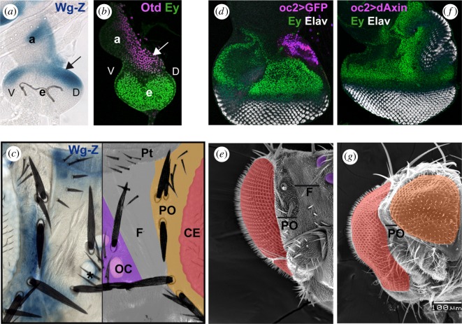
Attenuating the wg/Wnt signalling pathway results in the medial to lateral transformation of the Drosophila head. (a,b) Early third larva stage discs. (a) X-gal staining of the wg transcriptional reporter wg-Z. (b) Disc co-stained with anti-Otd (magenta) and anti-Ey (green) antibodies, showing complementary expression domains. The arrow in (a) and (b) points to the prospective dorsal head region, where ocelli develop. (c) X-Gal-stained wg-Z adult head (left) and schematic representation of dorsal head regions (right). wg expression is detected in the periocular cuticle (PO) and the anterior region of the dorsal head (ptillinum, PT). CE, compound eye; OC, ocellar complex; F, frons. The asterisk marks a late-appearing wg expression domain around the ocelli (see also [11]). (d,f) Late discs from oc2-GAL4; UAS-GFP (oc2 > GFP; d) or oc2-GAL4; UAS-dAxin2.28 (oc2 > dAxin; f), stained for the retinal marker Elav (white) and Ey (green). GFP expression in (d) is shown in magenta and marks the oc2-GAL4 expression domain. In oc2 > Axin discs, a duplicated eye field arises from the ocellar domain. (e,g) SEM images of oc2 > GFP (e) and oc2 > Axin adult half-heads shown at the same magnification. CEs are pseudocoloured in red. Ocelli in (e) are pseudocoloured in purple and the ectopic eye in (g) in orange. PO and F as in (c).

