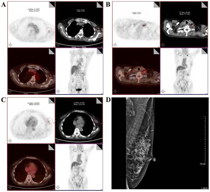
Figure 1.
18F-Fluorodeoxyglucose (FDG) positron emission tomography/computed tomography (PET/CT) scan and the mammogram of the patient. (A) 18F-FDG-PET/CT scan showing intense hypermetabolism at the level of the enlarged left axillary lymph nodes, with a maximum standardized uptake value (SUVmax) of 3.6. (B) Hypermetabolism at the level of the enlarged left subclavian lymph nodes (SUVmax, 4.9). (C) Lesion with uneven density in the lower outer quadrant of the left breast (SUVmax, 2.0). (D) Mammography revealed the presence of a lobulated high-density mass in the left breast. The red circles indicate the lesion.

