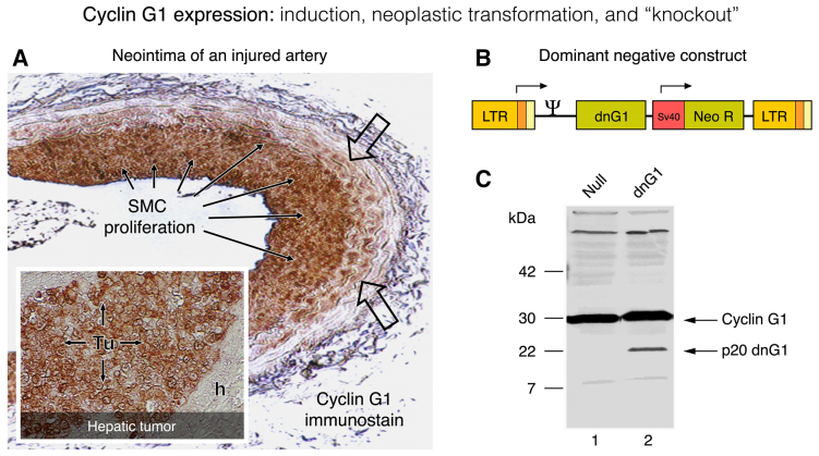
Figure 4.
Cyclin G1 expression: Induction, neoplastic transformation and knockout. The characteristic induction of cyclin G1 protein (G0-to-G1 phase) is visualized by immunohistochemistry (A; brown staining) in the activated periphery (open arrows) and the proliferative smooth muscle cells (SMC; solid arrows) in a rat carotid artery model of vascular restenosis following balloon catheter injury. For comparison, the constitutively high levels of cyclin G1 expression seen in a flagrant pancreatic cancer xenograft (Tu), metastatic to the liver (A; insert) is contrasted by the negligible expression of cyclin G1 seen in the adjacent normal (host) nude mouse hepatocytes(h). The design of a dominant-negative (knockout) construct of cyclin G1 (p20 dnG1) is shown in the context of its MoMuLV retroviral expression vector (B); the truncated p20 dnG1 protein (devoid of N-terminal and α1, α2 cyclin box domains) induces apoptosis in the presence of the abundant wild-type cyclin G1, as shown in western blots of cellular proteins (C).

