Figure 7.
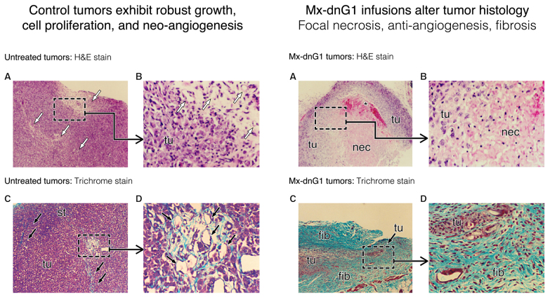
Characteristic changes in tumor histology following intravenous Mx-dnG1 treatment. Untreated tumor xenografts (left panel) exhibit robust tumor (tu) cell proliferation with active zones of neo-angiogenesis (A, open arrows; B, magnified); Masson's trichrome stain (C, D magnified) reveals exposed collagenous proteins (solid arrows, blue stain) associated primarily with angiogenesis and reactive stroma formation (st). By contrast, Mx-dnG1 treatment (right panel) reduces the flagrant population of proliferative tumor cells (tu) significantly (A, boxed; B, magnified), producing large regions of focal necrosis (nec) accompanied by overt anti-angiogenesis (*). Residual tumors are ultimately reduced to besieged clusters of proliferative tumor (tu) cells (C, boxed; D, magnified), as reactive/reparative fibrosis (fib), including nascent (i.e., targetable) ECM deposition (blue stain), dominates the histology of tumor regression observed in Mx-dnG1 treated animals.

