Fig. 3.

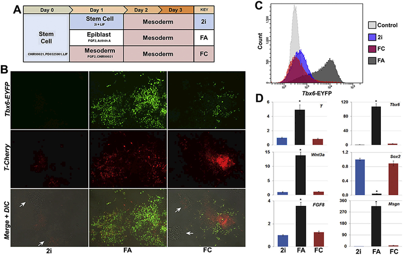
Conversion of mouse ESCs to epiblast state enhances paraxial mesoderm differentiation. (A) Schematic depiction of differentiation conditons. Note day 1 conditions for comparing delayed (2i), direct (FC), and epiblast (FA) transitions into mesoderm. (B) Representative images of reporter expression on day 3 of differentiation showing robust Tbx6 reporter expression and spreading of T+ cells following epiblast (FA) transition. T+ cells remained clustered following direct mesoderm induction (FC), with limited Tbx6 reporter expression. The persistence of numerous mounded, reporter-negative colonies occurred with the 2i and FC transitions (white arrows), which were absent from epiblast-transitioned cultures. (C) Day 3 FACS analysis shows epiblast transition (FA) followed by two days of mesoderm differentiation generated considerably more Tbx6+ cells compared to just two (2i) or three (FC) days of mesoderm differentiation, with control stem cells shown for comparison. (D) Day 3 gene expression indicated elevated levels of early mesoderm genes T, Wnt3a, and Fgf8 and substantial increases in the paraxial markers Tbx6 and Msgn with FA transition compared to 2i or FC, which also retained expression Sox2. Data shown as mean ± SEM (n = 3, *p < .05, where * denotes the comparison of FA treatment to both 2i and FC).
