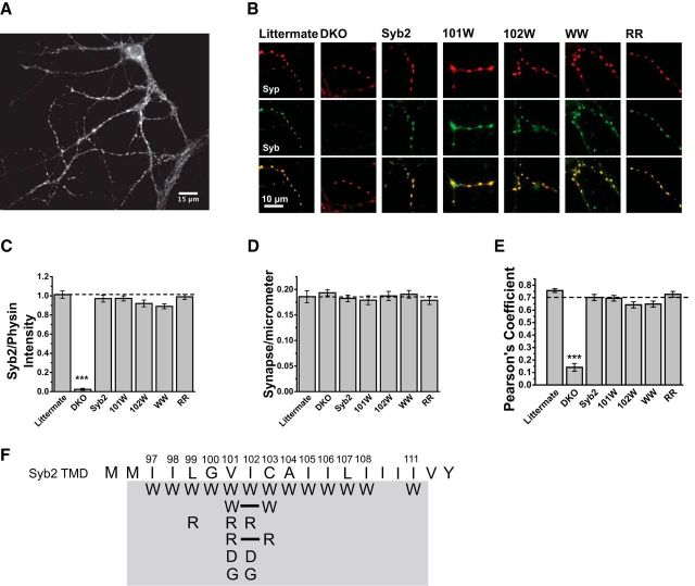
Figure 1.
Expression of WT Syb2 and TMD mutants in dissociated Syb2/cellubrevin DKO hippocampal neurons. A, Syb2 immunofluorescence reveals synaptic boutons on the dendrites of a cultured neuron. B, Double immunofluorescence of dendritic segments with synaptic boutons show the endogenous synaptic vesicle marker synaptophysin (top, red), Syb2 (middle, green), and the two images merged (bottom), in littermate control neurons, untransfected neurons, and neurons expressing WT Syb2 or four selected TMD mutants, V101W, I102W, V101W/C103W (WW), and V101R/C103R (RR). C, The fluorescence intensity of puncta in Syb2 images such as those in A and B were compared. Intensities are indistinguishable between littermate neurons, DKO cells expressing WT Syb2, and DKO cells expressing the selected Syb2 mutants. The puncta intensity in DKO (N = 19) was lower than littermate (N = 11; two-tailed Mann–Whitney test, U = 209, p = 7.6 × 10⁻⁶) and WT Syb2 (N = 19; U = 361, p = 1.5 × 10⁻⁷). All values were relative to synaptophysin (physin) intensity of the same image. D, The number of synapses per micrometer of dendrite, determined by synaptophysin immunofluorescence, was indistinguishable between all conditions examined using one-way ANOVA with the Bonferroni post hoc test. E, Colocalization between endogenous synaptophysin and overexpressed Syb2 was evaluated with Pearson's correlation coefficient, and was indistinguishable between Syb2 expressing neurons by one-way ANOVA with the Bonferroni post hoc test, but lower in untransfected DKO neurons. F, Schematic of Syb2 TMD displaying all the mutants (gray background) tested in this study. Solid lines indicate double amino acid substitutions. ***p < 0.001.

