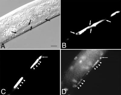
Figure 2.
rho-1(dn) can cause a cytokinesis defect. (A) Abnormally large neurons (flanked by arrows) in a living rho-1(dn) animal during the second larval stage. (B) Fluorescent visualization of the unc-47∷GFP-positive cells (flanked by arrows) in the animal shown in A. (C) Fluorescent visualization of unc-47∷GFP-positive neurons in a fixed rho-1(dn) animal. (D) DAPI visualization of the multiple nuclei observed in the unc-47∷GFP-positive cells shown in C. Arrowheads in C and D denote the positions of nuclei as determined by DAPI staining. Arrows in C and D show an unc-47∷GFP-positive cell without the cytokinesis defect. (Bar = 7 μm.) In wild-type animals (see Fig. 1A), multiple neurons are clearly visible in the same region of a multinuclear cell in the mutant.

