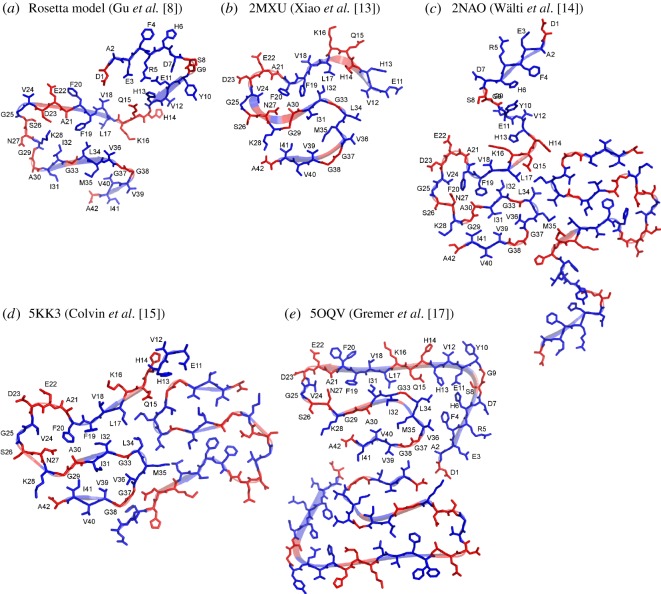
Figure 3.
Comparison of Aβ42 fibril models in the context of site-specific structural order as determined from EPR data. The structural model based on EPR data and Rosetta modelling is shown in panel (a). Four recent structural models of Aβ42 fibrils from the Protein Data Bank are shown in panels (b)–(e). The secondary structure is shown as ribbons, and the information on the secondary structure is taken directly from PDB files. Residues with strong spin exchange interactions are shown in blue, and residues with weak spin exchange interactions are shown in red.

