
Figure 3. Nucleocytoplasmic transport factors are constituents of stress granules.
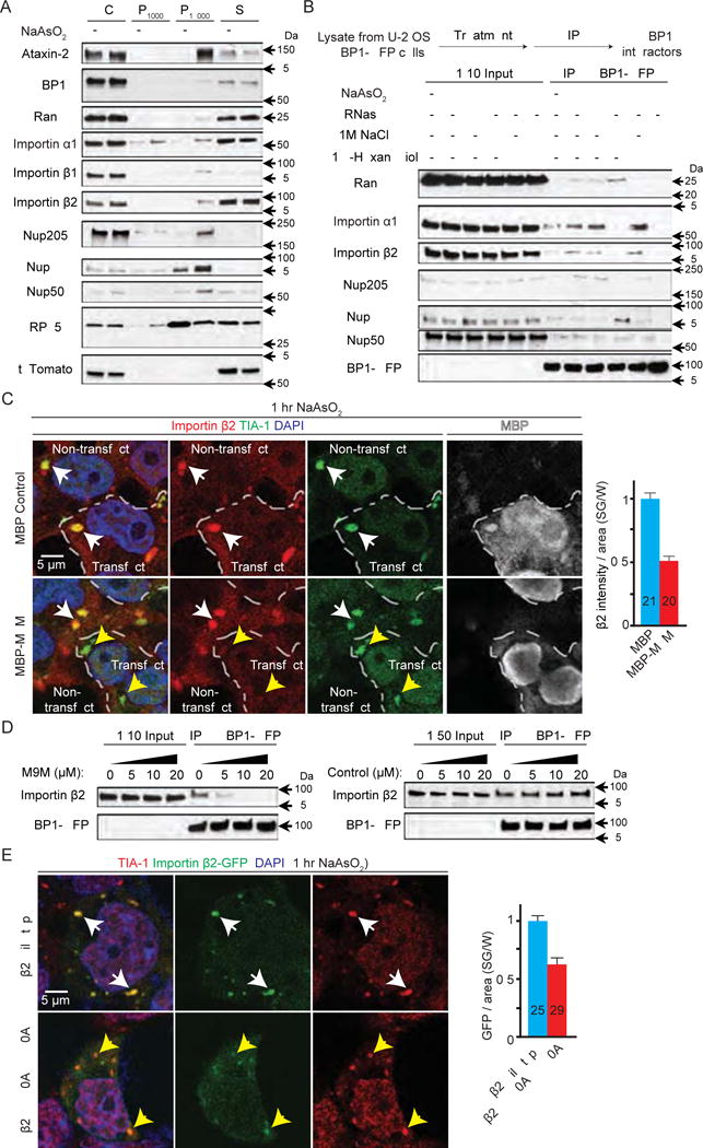
(A) Subcellular fractionation of HEK293T cells. P1000: pellet from 1,000 g; P18000: pellet from 18,000 g; S: supernatant after 18,000 g. (B) Co-IP of nucleocytoplasmic transport factors with G3BP1-GFP from U-2 OS cells expressing G3BP1-GFP. (C) HEK293T cells expressing MBP (control, top) or MBP-tagged M9M (bottom) were stained with Importin β2 (red), TIA-1 (green), DAPI (blue), and MBP (white). Dashed lines separate transfected versus non-transfected cells. White arrowheads indicate co-localization. Yellow arrowheads indicate TIA-1-positive puncta without Importin β2 co-localization. (D) Co-IP of Importin β2 and G3BP1-GFP with chemically synthesized M9M or control peptide. (E) HEK293T cells expressing GFP-tagged wild type (top) or mutant (bottom) Importin β2 (green) stained with TIA-1 (red) and DAPI (blue). Arrowheads indicate co-localization. W: whole cell, n numbers in graph. ****: p<0.0001. Data are represented as mean ± SEM.