
Figure 6. The N- and C-termini of MRI are both required for its function in cNHEJ.
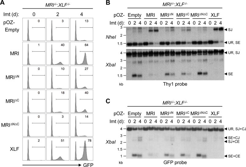
(A) Flow cytometric analyses of GFP expression in MRI−/−:XLF−/− abl pre-B cells retrovirally transduced with MRI, MRIΔN, MRIΔC, MRIΔNΔC, or XLF and treated with imatinib for the indicated lengths of time (days, d). (B) Southern blot analyses of genomic DNA from cells in (A) that were digested with NheI (top) or XbaI (bottom) and hybridized to the Thy1 probe. (C) Southern blot analyses of genomic DNA from cells in (A) that were digested with XbaI and hybridized to the GFP probe. Bands corresponding to different pMG-INV arrangements are indicated as described in Figure 3.