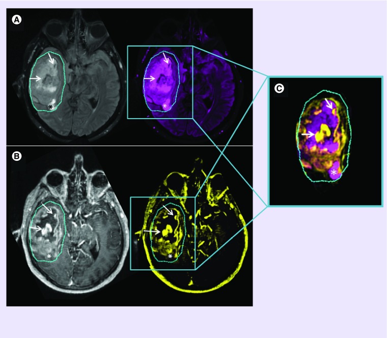
Figure 1. . Conventional MRI.
T2-weighted FLAIR images (A) are used to non-specifically assess edema surrounding the T1-weighted contrast-enhancement (B). Though signal hyper- or hypo-intensities may indicate certain biological scenarios, one cannot be certain of the eco-biology within ROIs. Combined ROIs (C) of low T2-FLAIR with high T1-post-contrast signal (arrows) can imply absence of edema, as a result of tumor and/or inflammatory cell presence, with compromised BBB integrity and leaky vasculature; ROIs of increased T2-FLAIR with low T1-post contrast signal (*) can imply regional edema, resulting from an adjacent inflammatory response or from an absence of tumor cells (possible necrosis), with a probable intact BBB and preserved vascular integrity. Contrary to what may be expected, a low signal on a T1-post-contrast image does not necessarily imply a reduced or compromised blood supply- information of this nature would only be obtainable with perfusion imaging.
BBB: Blood–brain barrier; FLAIR: Fluid attenuated inversion recovery; ROI: Region of interest.

