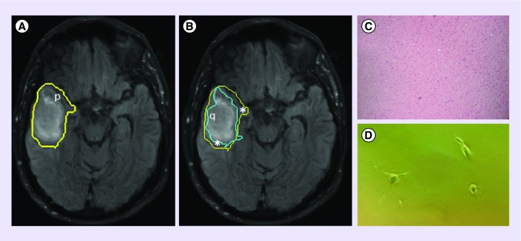
Figure 2. . T2-weighted FLAIR image of a GBM lesion with a superimposed delineation of the p-abnormality, which extends beyond FLAIR enhancement (A).
This, in conjunction with the q-abnormality (B), can be used to differentiate the tumor bulk (q) from the margin (*), which has a normal †histological appearance (C), but which harbors tumor ††cells (D). Regions of p- and q- abnormality were obtained using the p- and q- components of the diffusion tensor, as described by [30].
†Tissue samples obtained using 5-ALA fluorescence-guided resection, allowing access to normal-appearing tissue, stained with H&E, 10× magnification.
††Margin cells were plated in vitro and observed 4DIV.
5-ALA: 5-aminolevulinic acid; DIV: Days in vitro; FLAIR: Fluid-attenuated inversion recovery; GBM: Glioblastoma; H&E: Hematoxylin and eosin stain.

