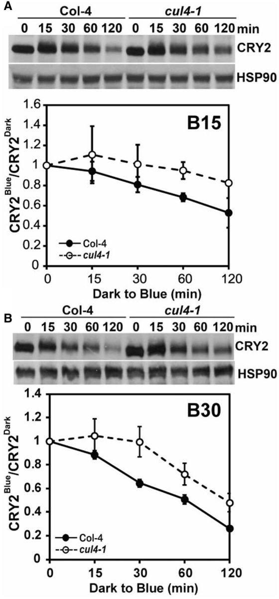
Fig. 3.

Cullin 4 is involved in blue light-dependent CRY2 degradation. (A) Immunoblot showing blue light-dependent CRY2 degradation in the cul4-1 mutant. Three-week-old wild-type (Col-4) and cul4-1 mutant plants grown in LD were adapted to dark for 24 h and treated with blue light (15 μmol m-2 s-1) for the times indicated before sample collection. The immunoblots were probed with anti-CRY2 antibody (CRY2) and reprobed with anti-HSP90 as the loading control. The relative band intensities of CRY2 were quantified and are shown with the SDs (n = 3). CRY2Blue, normalized (against HSP90) band intensity of CRY2 of blue light-treated samples, CRY2Dark, normalized band intensity of CRY2 of dark-treated samples. (B) Same as (A), except that plants were treated with blue light of 30 μμmol m-2 s-1. Three experiments were performed; one representative immunoblot and the relative levels of CRY2 (CRY2Blue/CRY2Dark) are shown with the SDs (n = 3).
