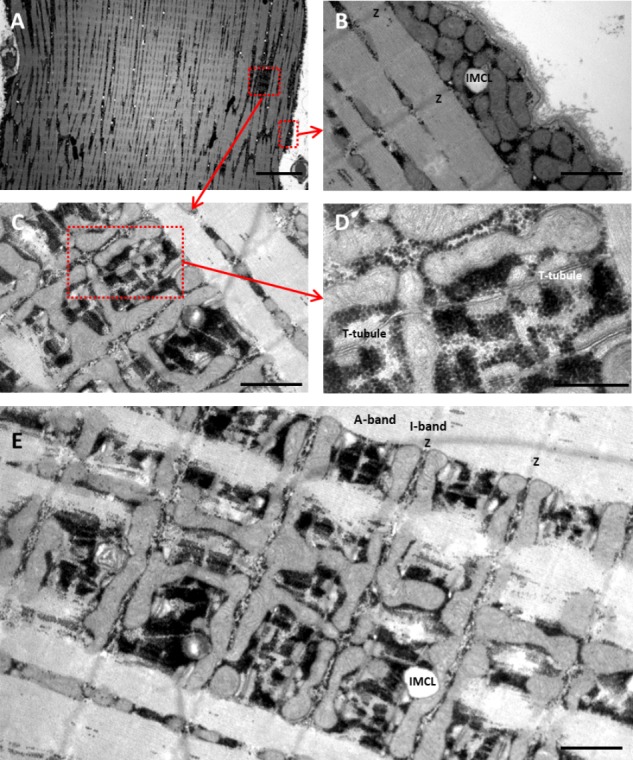
FIGURE 1.

TEM images showing the subcellular localization of skeletal muscle mitochondria. All images are from leg muscle (vastus lateralis). (A) Overview of a part of fiber showing the myofibrillar (Myo) space and subsarcolemmal (SS) space. (B) The typical localization of SS mitochondria (mit) in skeletal muscle, also showing a intramyocellular lipid (IMCL). (C) In the Myo space, intermyofibrillar mitochondria are wrapped around the myofibrils, mainly in the I-band and often connected to an adjacent mitochondrion through the A-band. There is less marked connection between neighboring mitochondria in the I-band. (D) Intermyofibrillar mitochondria in the I-band on each side of the Z-line, with the t-tubular system (t-system) and mitochondria intertwined. (E) Overview demonstrating the IMF mitochondria are mainly located in the I-band on each side of the z-line and often connected to an adjacent mitochondrion in the same sarcomere through the A-band. All the gray structures in the fiber are mitochondria with slightly visible inner cristae. Glycogen granules can be seen as black dots. Z, Z-line; A, A-band; IMCL, intramyocellular lipid; T-tubule, transverse tubular system. Scale bar: A, 10 μm; B,C, 1 μm; D, 0.5 μm; E, 1 μm. Original magnification: A, x1,600; B,C, x20,000; D, x50,000; E, x13,000.
