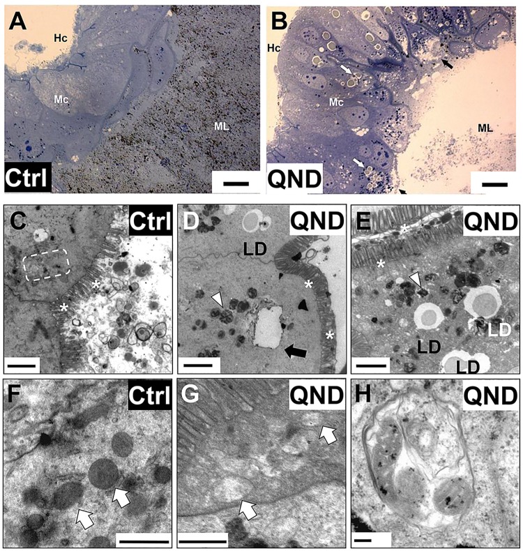
Fig 2. Impaired heme crystallization affects posterior midgut organization.
(A and B) Light microscopy images of posterior midguts of adult insects fed with blood (Ctrl; A) and blood supplemented with 100 μM QND (B) from IBqM colony four days after blood meal (bars = 20 μm). Midgut lumen (ML), midgut cells (Mc), hemocoel (Hc), vacuoles (black arrows) and lipid droplets (white arrows) are indicated in the images. (C-H) Transmission electron microscopy images of posterior midguts from insects maintained at IBqM (C-F), and Fiocruz (G,H) colonies fed with blood (Ctrl, C,F), or blood + 100 μM QND (D,E,G,H) four days after blood meal. The general architecture of posterior midgut cells in control insects (C,F) includes mitochondria (white arrows), endoplasmic reticulum (dashed box) and microvilli (asterisks). In QND treated insects (D,E,G,H), extensive organelle disappearance contrasts with the presence of numerous electron-dense hemoxisomes/residual bodies (arrowheads), vacuoles (black arrow), and intracellular lipid droplets (LD). Electron-dense mitochondria found in the posterior midgut of control insects (F), contrast with swollen and washed out mitochondria from 100 μM QND treated insects (G). (H) Mitochondria inside an autophagosome. (Scale bars: C-E: 2 μm; F: 1.0 μm; G,H: 0.5 μm).

