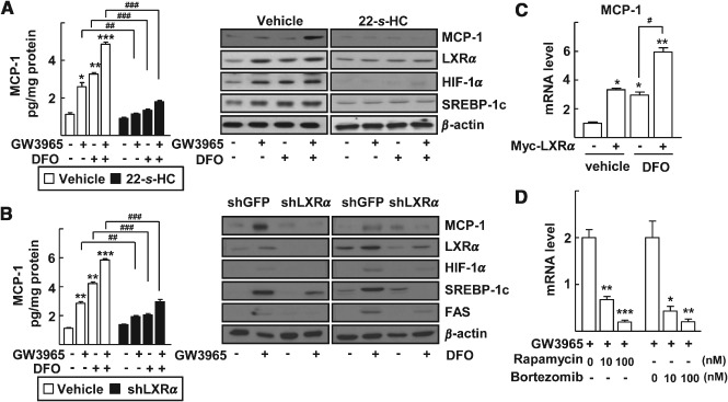
Figure 2.

Induction of MCP‐1 by LXRα and HIF‐1α in primary mouse hepatocytes. (A) Primary hepatocytes were treated with the indicated combinations of 1 µm GW3965, 100 µm DFO and 1 µm 22‐S‐HC for 24 h. (B) Primary hepatocytes were infected by lenti‐shGFP or lenti‐shLXRα viruses. After 24 h of viral infection, the cells were treated with the indicated combinations of 1 µm GW3965 and 100 µm DFO for an additional 24 h. MCP‐1 secreted into the culture supernatants was quantified by ELISA (left). The numbers represent mean ± SD (n = 3); *p < 0.05, **p < 0.01 and ***p < 0.001 compared with no treatment; ## p < 0.01 and ### p < 0.001 compared with vehicle or shGFP, as indicated: expressions of protein levels in whole‐cell lysates were analysed by western blotting (right). (C) Huh‐7 cells were transfected with empty vector or Myc‐LXRα; after 24 h, the cells were treated with vehicle or 100 µm DFO; mRNA levels of MCP‐1 were measured by qRT–PCR; numbers represent mean ± SD (n = 3); *p < 0.05 and **p < 0.01 compared with empty vector transfection and vehicle treated control; ## p < 0.01 and ### p < 0.001 compared with the treatment of DFO. (D) Huh‐7 cells were treated with 1 µm GW3965 in the presence or absence of rapamycin or bortezomib for 24 h; mRNA levels of MCP‐1 were measured by qRT–PCR; numbers represent mean ± SD (n = 3); *p < 0.05, **p < 0.01 and ***p < 0.001 compared with GW3965 alone; statistical significance was evaluated by two‐way ANOVA
