Figure 7.

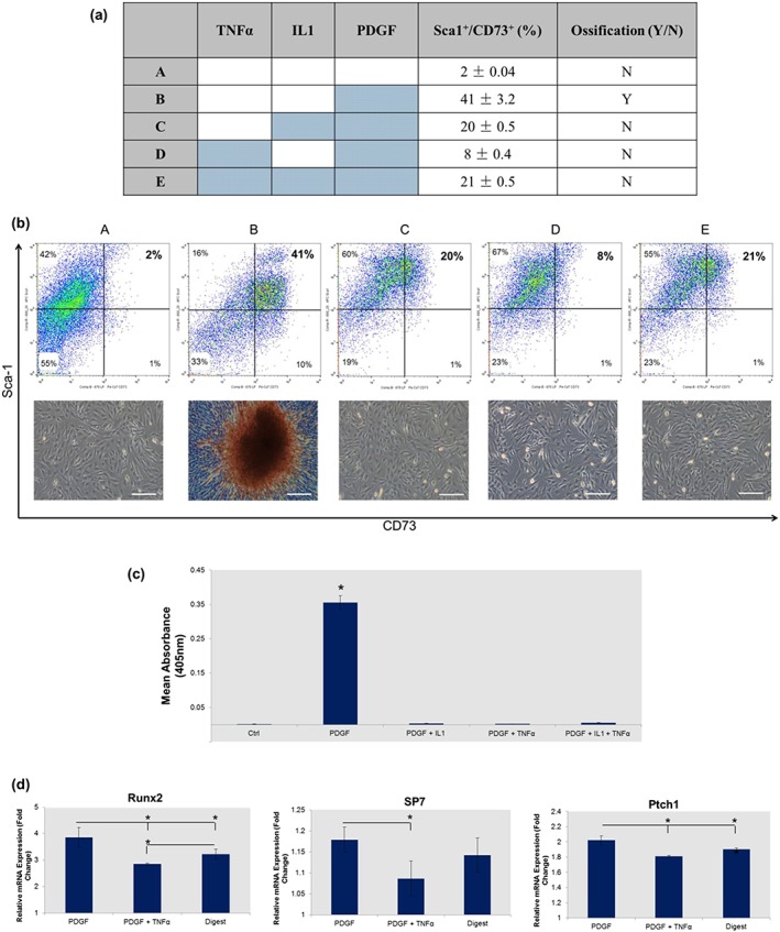
Pro‐inflammatory cytokines tumour necrosis factor (TNF)α and interleukin (IL)1 mediate platelet derived growth factor (PDGF)‐induced ectopic ossification in tissue‐engineered skeletal muscle. (a) Table displaying the combination of factors added to each culture and how this affected the proportion of Sca‐1+/CD73+ cells and the presence of ectopic mineral. (b) Flow cytometry data and Alizarin red staining showing the mediating effects of pro‐inflammatory cytokines on mineralization, and how this is related to the proportion of Sca‐1+/CD73+ cells. (c) Quantification of the concentration of Alizarin red stain bound in PDGF‐BB cultures exposed to pro‐inflammatory cytokines IL1 and TNFα. (d) Quantitative real‐time reverse‐transcription polymerase chain reaction gene expression analysis showing reductions in osteogenic regulators Runx2 and SP7, and the downstream hedgehog signalling factor Ptch1 in the presence of TNFα. Polymerase chain reaction was performed at day 3 with gene expression normalized to the untreated ‘reserve’ cells using the ΔΔCT (Livak) method. Coloured cells signify the addition of growth/inflammatory factors. Bars: 100 μm. *p < 0.05. Flow cytometry, polymerase chain reaction and Alizarin red quantification data are represented as mean ± standard deviation (n = 3)
